Ultra-high molecular weight polyethylene wear particle-induced osteolysis and, ultimately, implant loosening are recognized as major factors limiting the longevity of total joint replacement components. Highly crosslinked polyethylenes (HXLPE) were introduced to improve implant wear resistance, and have yielded improved outcomes with regard to the reduction of osteolysis at short-term and mid-term followup. However, there remains a considerable interest in understanding the characteristics, biological relevance and long-term effects of wear debris generated from newer, HXLPE liners in vivo.
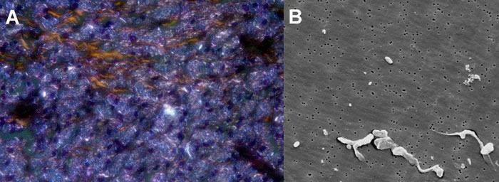
Methodologically, micron- and submicron-sized polyethylene wear debris can be observed in periprosthetic tissues using polarized light microscopy. In this approach, polarized light filters induce an intrinsic particle birefringence due to the anisotropy of the polymer structure (Figure A). Alternatively, using scanning electron microscopy, the size and morphology of nanometer- and submicron-sized polyethylene wear debris can be obtained in greater detail (Figure B, filter with a pore size of 50 nm is shown).